Each year the NDIIF recognizes excellence in imaging through publication as well as art. Year after year our students continue to impress us with their hardwork, diligence, and research efforts. Three awards are granted each year. The awards are judged on the basis of a piece of original research in the physical and material sciences involving electron microscopy as well as research with biological applications highlighting optical microscopy or InVivo Imaging. The selection of the winner takes into account the impact level of the journal, the cutting-edge discovery of the research conducted within the publication, and all levels of innovation and sophistication required to obtain the image. Collaboration across disciplines is encouraged but not a requirement for submission. Consideration is given to publications whose work utilized multiple facets within the NDIIF.
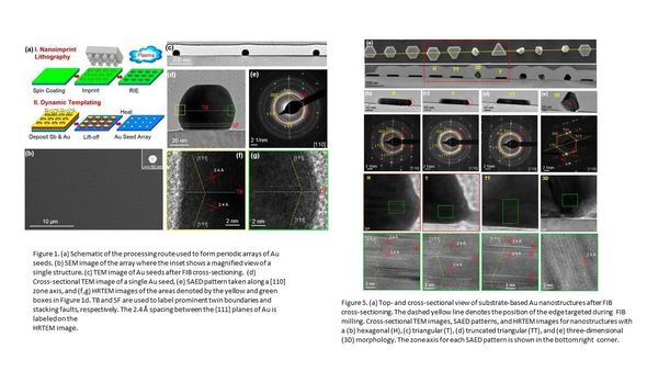
The winner of the Best Electron Beam Publication of 2019 is awarded to Spencer Golze for his work regarding Plasmon-Mediated Synthesis of Periodic Arrays of Gold Nanoplates Using Substrate-Immobilized Seeds Lined with Planar Defects published in Nano Letters, 201919, 5653-5660. The growth mechanisms and defect formation in Au seeds and nanoplates have been studied based on SEM and TEM analysis. A combination of High Resolution TEM and STEM imaging as well as Selected Area Electron Diffraction (SAED) was performed using an FEI Titan 80-300. TEM has been employed for structural and compositional characterization of the samples. TEM cross-sectional samples were prepared using an FEI Helios SEM/FIB dual beam tool. The cross-sectional TEM images provide evidence that Au seeds and the hexagonal nanoplates contain planar defects, twins, and stacking faults, that are parallel to the substrate surface. The presence of twins in the seeds and nanoplates is also evidenced by the corresponding electron diffraction pattern.
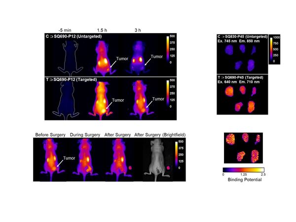
The winner of the Best Biological Imaging Publication of 2019 is awarded to Cynthia Spires for her work regarding Paired Agent Fluorescence Imaging of Cancer in a Living Mouse Using Pre-assembled Squaraine Molecular Probes with Emission Wavelengths of 690 and 830nm in Bioconjugate Chemistry, 2019, 31, 214-223. New imaging methods were evaluated with targeted fluorescence probes for imaging cancer in a living mouse model. First, Single Agent Imaging (SAI) experiments compared tumor imaging performance of a targeted probe and untargeted probe in separate mouse cohorts. Subsequently, a mock surgery was performed that completely removed the fluorescently labeled tumor. Furthermore, there was imaging evidence for enhanced tumor accumulation of the targeted probe, but there was moderate scatter in the data due to tumor-to-tumor variability. A subsequent Paired Agent Imaging (PAI) study co-injected a binary mixture of targeted probe (with emission at 690 nm) and untargeted probe (with emission at 830 nm) into the same tumor-burdened animal. The pairing provides a correction factor that accounts for tumor-to-tumor variability in nonspecific probe uptake with statistically higher tumor uptake of the targeted probe using a much smaller cohort of mice. The imaging data from the PAI experiment was analyzed to determine the targeted probe’s Binding Potential (BP) for available integrin receptors within the tumor tissue. In addition, pixelated maps of BP within each tumor indicated a heterogenous spatial distribution of BP values. PAI is a promising new way to evaluate targeted fluorescent probes and eventual use in clinical applications.
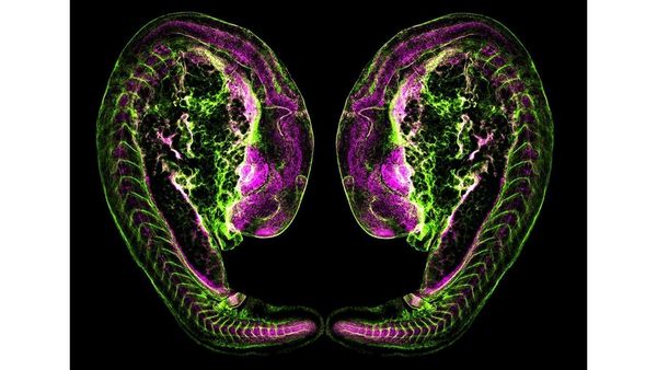
The winner of Best Artistic Image of 2019 is awarded to Brooke Chambers. This image was captured using the Nikon C2 confocal Microscope in our Optical Microscopy Core. Featured is a reflected 10X image of a beautiful model organism, the Danio rerio embryo, commonly known as the zebrafish. At just 1 day post fertilization, the zebrafish has developed eye structures, a kidney tubule, and 28 chevron-shaped muscle units. Specifically, this whole mount immunofluorescent preparation labels laminin (basement membranes) in green and Slc12a1/2 expression (solute transporter proteins) in magenta. Because the embryonic zebrafish undergoes rapid organ formation and is optically transparent in nature, it is an ideal system for high resolution imaging of dynamic developmental processes. Although a simpler vertebrate species, the zebrafish exhibits striking genetic, cellular, and anatomical similarities to humans. This image brings to light the boundless potential of the zebrafish to model human disease and embryonic development.
Congratulations to Spencer, Cynthia, and Brooke as well as all of the outstanding nominations that the Imaging Facility received this year! We appreciate your hard work and your dedication to continue moving research forward.