Two researchers from the University of Notre Dame have received recognition for publications and images from the Notre Dame Integrated Imaging Facility (NDIIF). Each year, the NDIIF provides imaging awards to recognize and honor researchers who use the facility equipment in two categories: biological publication imaging and material sciences publication imaging.
“We are grateful to all of the users of the NDIIF. Our annual imaging awards provide us an important opportunity to recognize their discoveries,” said Bradley Smith, Emil T. Hofman Professor of Science in the Department of Chemistry and Biochemistry and director of the NDIIF. “This year’s awardees showcase the breadth and the beauty of the research taking place within the facility.”
Best Biological Publication Image
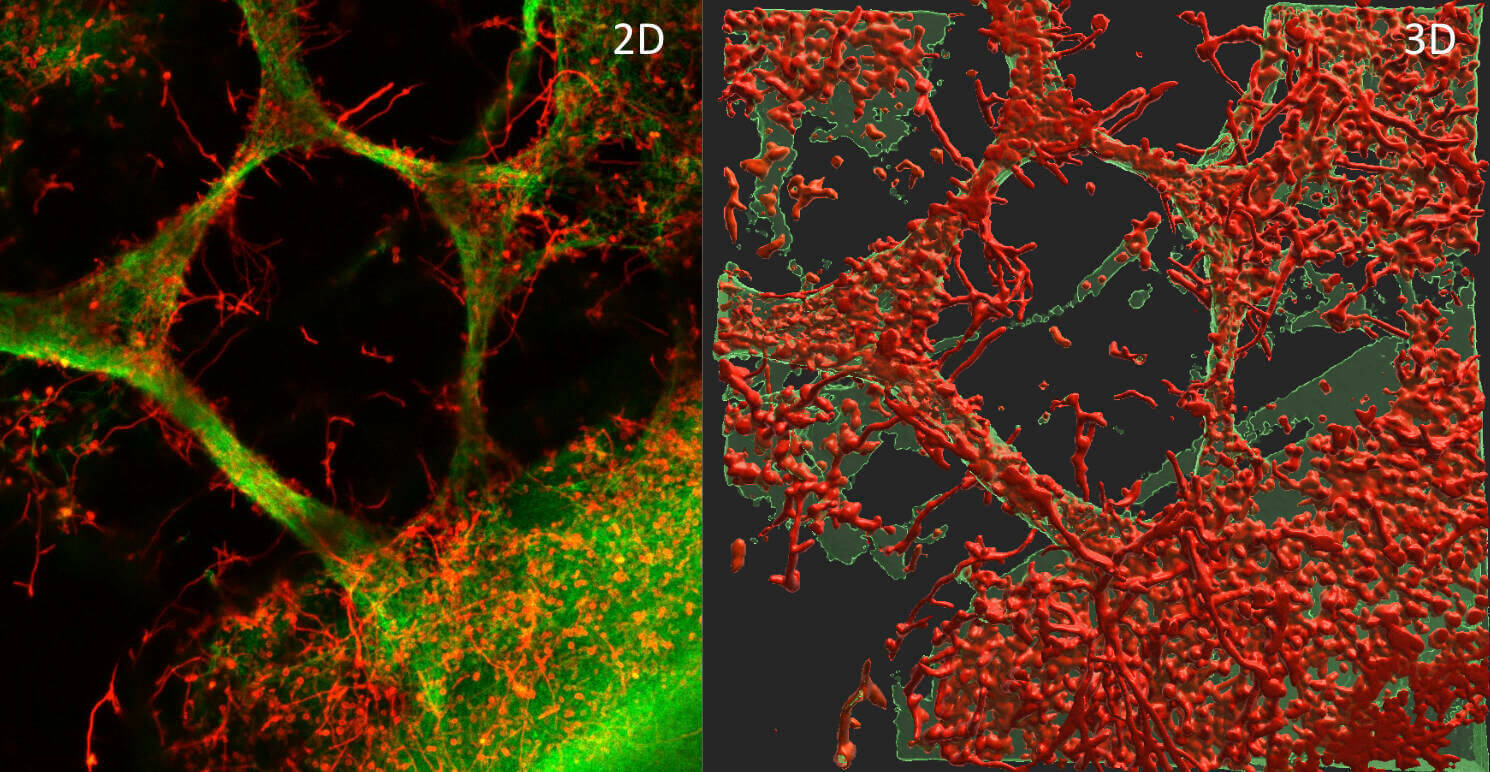
Alyssa LaBella, a doctoral student in the Department of Biological Sciences, received the award for best biological publication imaging. The winning image appeared in Science Advances in the article “The catheterized bladder environment promotes Efg1-and Als1-dependent Candida albicans infection.”
Catheter-associated urinary tract infections (CAUTIs) are the most common healthcare-associated infection. (More than 150 million individuals are infected each year.) The researchers offered a deeper understanding of how catheterization affects the bladder and why it creates an environment where Candida albicans infections occur and persist.
LaBella is a member of the Flores-Mireles Lab, led by Ana Lidia Flores-Mireles, the Janet C. and Jeffrey A. Hawk Assistant Professor of Biological Sciences. LaBella is also mentored by Felipe H. Santiago-Tirado, an assistant professor in the Department of Biological Sciences who is one of the paper's co-authors.
Best Material Sciences Publication Image
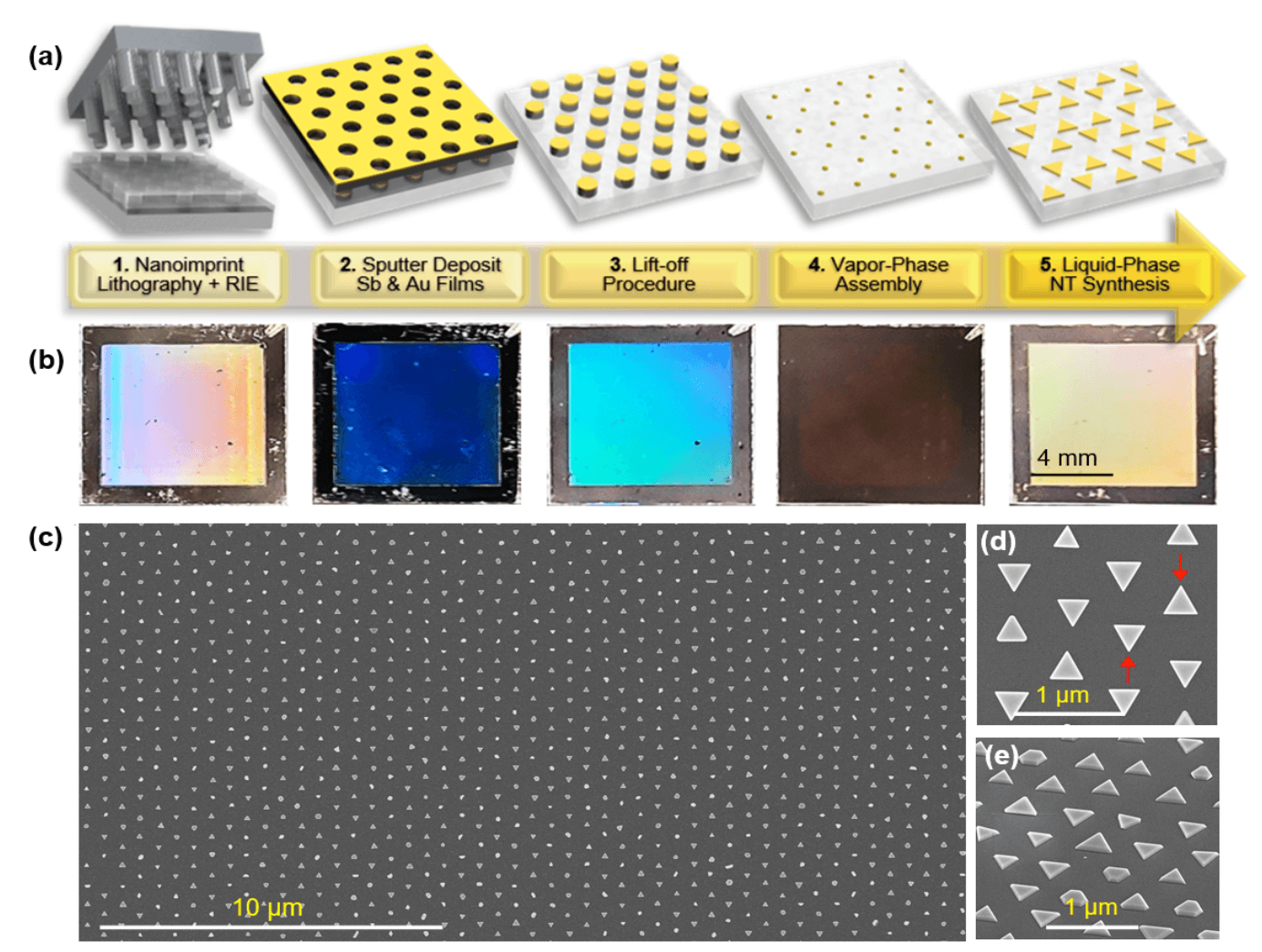
Robert D. Neal, adjunct assistant professor in the Department of Aerospace and Mechanical Engineering, received the award for best material sciences publication image for research published in Small, entitled “Large-area periodic arrays of atomically flat single-crystal gold nanotriangles formed directly on substrate surfaces.”
The paper describes a simple and cost-effective five-step procedure for fabricating plasmonic gold nanotriangles on substrates. This newly developed process demonstrated the first ever procedure capable of growing these nanotriangles in oriented arrays directly on substrate surfaces, and could prove useful in designing and manufacturing the next generation of electronic devices.
Neal worked alongside Svetlana Neretina, a professor in the Department of Aerospace and Mechanical Engineering.
All awarded images were taken on NDIIF equipment and were taken by a Notre Dame faculty, staff, or student. Both of these accomplishments were recognized at the 8th Annual Midwest Microscopy and Microanalysis Workshop on May 11, 2023.
The NDIIF at the University of Notre Dame provides an integrated suite of sophisticated microscopes and imaging stations that enable expert users to attack the most complex research problems as well as resident professional staff, including technicians and research specialists, to guide non-expert users. The facility is open to campus and external users. To learn more about the facility, please visit imaging.nd.edu.
Contact:
Brett Beasley / Writer and Editorial Program Manager
Notre Dame Research / University of Notre Dame
bbeasle1@nd.edu / 574.631.8183
research.nd.edu / @UNDResearch
About Notre Dame Research:
The University of Notre Dame is a private research and teaching university inspired by its Catholic mission. Located in South Bend, Indiana, its researchers are advancing human understanding through research, scholarship, education, and creative endeavor in order to be a repository for knowledge and a powerful means for doing good in the world. For more information, please see research.nd.edu or @UNDResearch.
Originally published by at research.nd.edu on May 22, 2023.