Multiplex Immunohistochemistry (mIHC) stands at the forefront of contemporary biological and medical research, revolutionizing the analysis of tissues and cells. It represents an evolution from traditional immunohistochemistry (IHC), allowing for the simultaneous examination of multiple proteins or antigens within a single tissue sample. While conventional IHC typically visualizes one protein or antigen at a time, mIHC utilizes a combination of specialized antibodies tagged with distinct fluorophores or chromogens. These tagged antibodies are applied to tissue sections, and advanced imaging techniques such as fluorescence microscopy or multispectral imaging enable the simultaneous visualization and analysis of various markers.
Visualization Techniques:

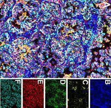
FIGURE 1: Illustrative mIHC/IF Images via Cutting-Edge Imaging Systems: The use of cutting-edge imaging systems—such as the Vectra, Chip cytometry, or DISCOVERY ULTRA—captures mIHC/IF images displaying a diverse array of labeled markers in tissue samples from different studies. For instance:
- (A) Pancreatic adenocarcinoma sections labeled with multiple markers via the Vectra imaging system.
- (B) Mouse pancreas sections labeled using the Chip cytometry imaging system.
- (C) Cholangiocarcinoma sections imaged through the DISCOVERY ULTRA system. These images exemplify the diverse applications and capabilities of mIHC/IF technology for various tissues and antigens.
Advantages:
Multiplex Immunohistochemistry offers several distinct advantages crucial for research and clinical applications:
-
Multiple Antigen Detection: Simultaneous visualization of multiple antigens within a single tissue sample.
-
Spatial Information: Understanding complex spatial relationships among proteins in tissues.
-
Reduced Sample Consumption: Maximizing the utility of limited samples.
-
Quantitative Analysis: Enables precise and quantitative data collection and analysis.
-
Enhanced Insights: Providing a deeper understanding of cellular interactions and tissue microenvironments.
Multiplex IHC Platforms:
Diverse platforms and methods have emerged to facilitate the comprehensive visualization and analysis of multiple proteins or antigens in tissue samples. These platforms vary in the number of markers they can detect simultaneously, spatial resolution, sensitivity, and user-friendliness. Considerations for choosing a platform include research needs, experimental complexity, sample type, imaging capabilities, and data analysis tools. Some prominent platforms include:
-
OPAL Multiplex IHC Kits (PerkinElmer): Enables labeling multiple targets with distinct fluorophores for visualization.
-
Akoya Biosciences CODEX: High-plex mIHC system utilizing DNA-barcoded antibodies for high spatial resolution and quantitative analysis.
-
NanoString GeoMx DSP: Designed for spatial genomics, offering simultaneous detection of multiple targets combined with gene expression analysis.
-
Fluidigm Imaging Mass Cytometry (IMC): Combines mass spectrometry with microscopy, allowing for the simultaneous detection of over 40 metal-labeled antibodies for multiplex imaging.
-
DISCOVERY ULTRA: An imaging system offering software solutions for the analysis of multiplexed IHC data, compatible with various imaging systems.
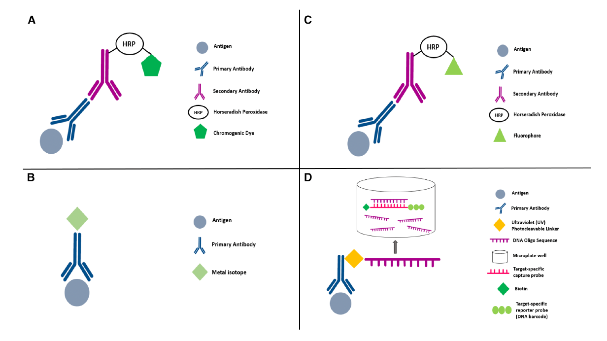
FIGURE 2: Mechanisms of mIHC/IF Platforms: The diagram displays the mechanisms of each mIHC/IF platform, showcasing their unique processes for antigen visualization and detection.
Multiplex Immunohistochemistry (mIHC) has emerged as a transformative force in modern biology and medical research, allowing for a comprehensive and simultaneous analysis of multiple proteins within tissue samples. By building upon the foundations of traditional immunohistochemistry, this innovative technique offers researchers a profound understanding of intricate cellular interactions and spatial relationships in tissues. Its advantages, including the detection of multiple antigens, provision of spatial information, reduced sample consumption, and enabling quantitative analysis, position mIHC as a crucial tool in fields such as cancer research, immunology, and beyond. With a diverse range of advanced platforms like OPAL Multiplex IHC Kits, Akoya Biosciences CODEX, NanoString GeoMx DSP, Fluidigm Imaging Mass Cytometry (IMC), and DISCOVERY ULTRA, scientists can choose tailored solutions for their specific research needs, ushering in a new era of comprehensive and in-depth cellular exploration and clinical applications.
The Notre Dame Integrated Imaging Facility is looking to add mIHC to one of the many services that the Histology Core has to offer. We would like to gather input and ask if you have an interest in mIHC that you please take a moment to answer a few brief questions. This will allow us to have a better understanding of what the needs of our University are and how we can better serve you. To answer the survey click here.
Or open the survey directly using a new tab https://nd.qualtrics.com/jfe/form/SV_3f2iRrVOYQgdzzU