Services
Supported by an expert team, the Notre Dame Integrated Imaging Facility (NDIIF) houses over twenty instruments with comprehensive capabilities to help answer many of the most relevant materials and biological questions researchers have today. Descriptions of each instrument along with their capabilities and rates can be viewed below.
For more information on using NDIIF instruments and services, including consultations and training, please visit the Use Our Services page.
Electron Microscopy Core
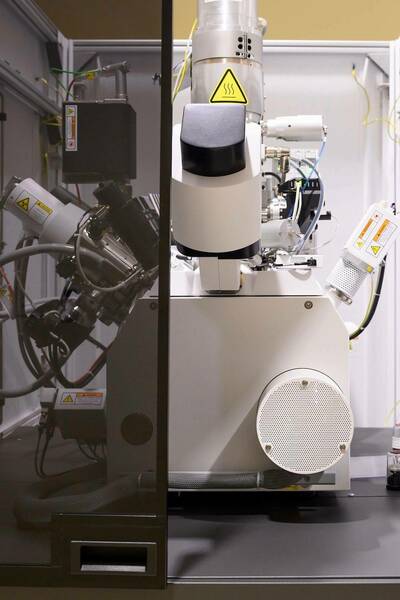
The Electron Microscopy Core integrates a unique bundle of state-of-the-art FEI tools that enable users to attack the most complex modern research problems from micron to Angstrom spatial resolution.
-
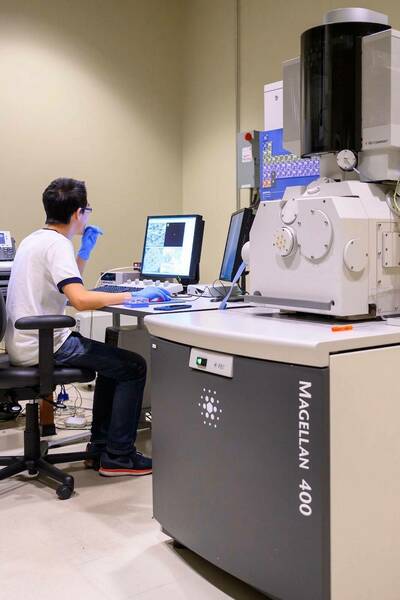
A field emission scanning electron microscope (FESEM) with extra high spatial resolution (XHR) which allows sample imaging at extremely low beam energies (<100 eV), avoiding charge effect at non-conductive nano-scale surfaces
-
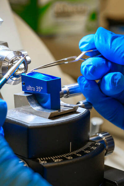
Designed for the highest-quality imaging, ultra-thin TEM and APT sample preparation, milling and deposition of complex structures with critical dimensions of less than 10 nm, multi-modal subsurface and 3D data collection
-
Spectra 30-300 (S)TEM – the highest resolution, aberration-corrected, scanning transmission electron microscope for all materials science applications
-
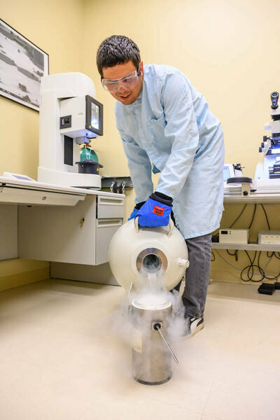
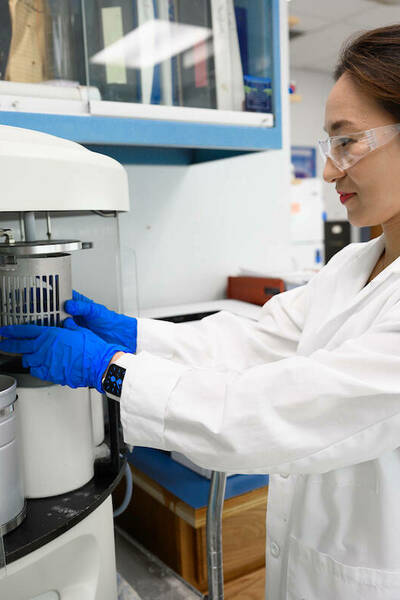
Leica Automatic Plunge Freezer EM GP2
For the preparation of vitrified fluid samples for cryo-TEM, including biological suspensions in both, aqueous and inorganic solvents
-
The Leica EM UC7 ultramicrotome equipped with the FC7 cryo chamber attachment is capable of preparation of high quality semi- and ultra-thin sections of biological and industrial samples
-
Talos F200i (S)TEM is a 20-200 kV field emission (scanning) transmission electron microscope uniquely designed for performance and productivity across a wide range of Materials Science samples and applications.
-
LYNX II Automated Tissue Processor
LYNX II is a batch tissue processor compatible with plastic resin and paraffin. LYNX II Automated Tissue Processor is the most unique state-of-the-art tissue processor. For EM and/or Histology. Easy programming. Internal battery back-up. Built-in fume extraction.
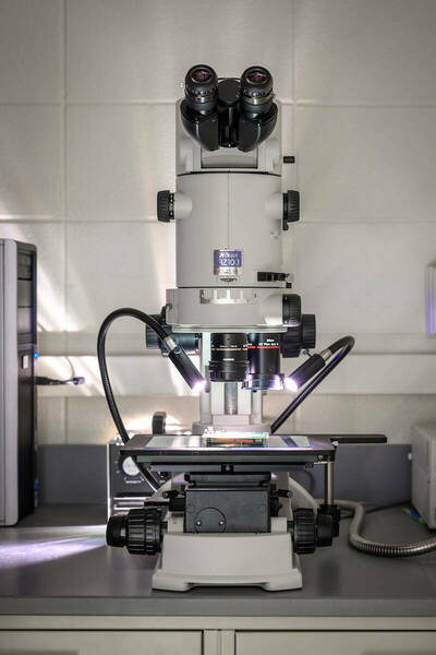
Optical Microscopy Core
The Optical Microscopy Core provides researchers with the opportunity to acquire informative single images and three-dimensional reconstructions of fluorescently labeled cells and tissues, either fixed or live.
-
The Nikon AZ100 is a multi-zoom macroscope with a wide range of magnification settings (5x-400x) used for either brightfield (top lit or backlit) or contrast imaging
-
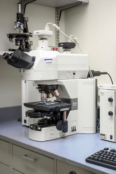
Nikon Eclipse 90i Widefield Fluorescent Microscope
The 90i is useful for imaging fluorescent or chromogenic (chemically stained) samples such as H&E, immunohistochemistry (i.e. DAB, HRP), Gram, trichrome, and more
-
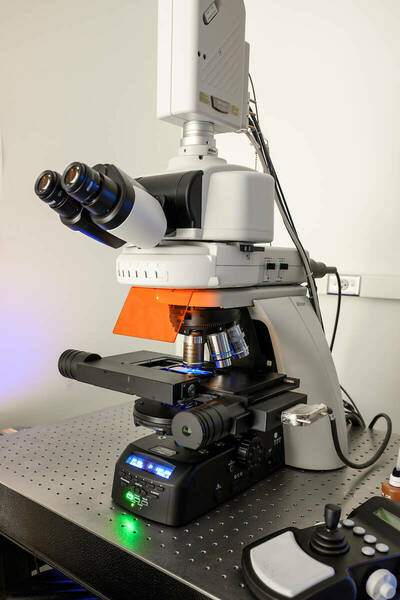
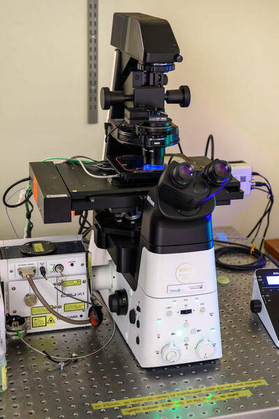
Nikon C2+ Laser Scanning Confocal Microscope
The Nikon C2 confocal system is a semi-automated, upright microscope ideal for imaging fixed samples providing basic confocal imaging with 3 standard PMT detectors and 4 laser lines for four channel imaging
-
The Nikon A1R inverted confocal system is equipped with four laser lines, 405, 488, 561 and 633nm and a contrast imaging capability and offers high-resolution imaging capabilities for a wide range of imaging applications
-
Nikon A1R-MP Laser Scanning Confocal Microscope
The two-photon imaging mode of the Nikon A1R-MP system provides deeper imaging into thick samples with no background resulting from out-of-focus light and minimal phototoxicity in live samples
-
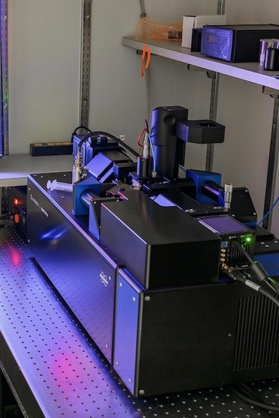
Bruker Luxendo MuVi SPIM Light Sheet
The lightsheet system is ideal for long-term, three-dimensional fluorescence imaging of organisms, tissue explants, and 3D cell culture systems. This microscope can be used for live, fixed, and optically cleared samples
-
Nikon CSU-X1 Spinning Disk System
The Nikon CSU-X1 is optimized for high speed imaging of living cells expressing fluorescent proteins and/or stained with synthetic fluorescent dyes
-
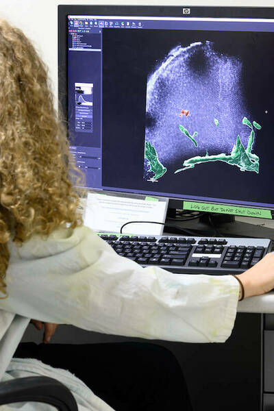
IMARIS software can be used for image presentation and/or data analysis from a variety of commercial fluorescence microscopy image file formats
-
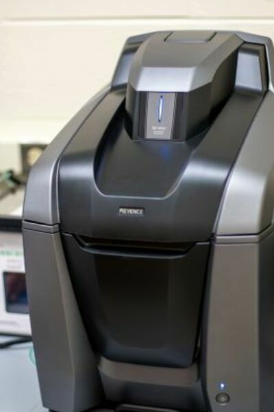
The Keyence BZ-X810 is a fully automated, brightfield and fluorescence microscope adaptable to many different research needs.
-
The multiphoton microscope STELLARIS 8 DIVE provides you with flexible multicolor imaging beyond 1 mm in depth.
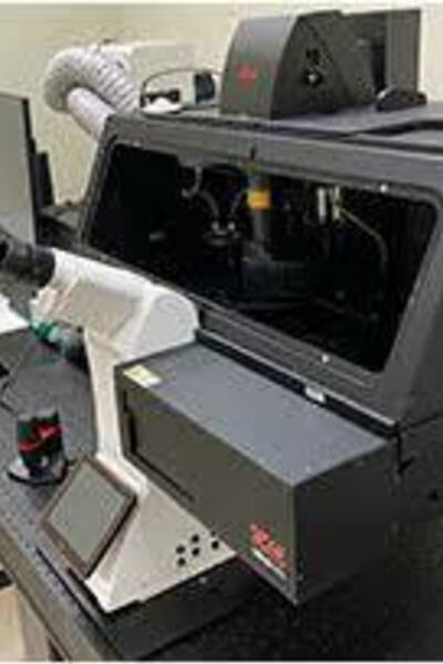
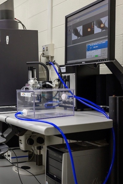
In Vivo Imaging Core
The Vivo Imaging Core provides a non-invasive approach to observe various disease and biological conditions in living systems.
-
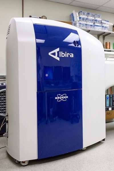
A small animal PET/SPECT/CT imaging system
-
A benchtop system for 2D bioluminscence, standard fluorescence, and Cerenkov imaging.
-
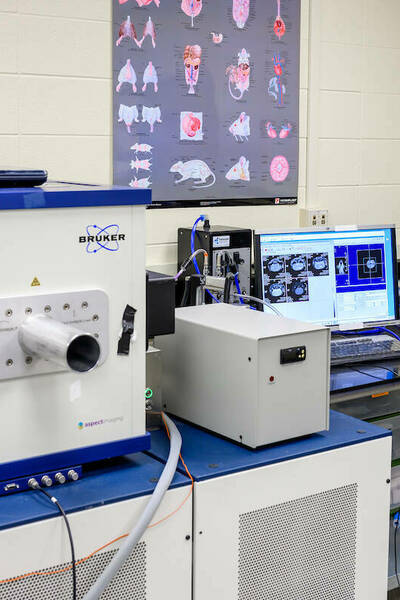
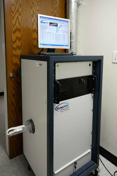
An easy-to-use 1 Tesla desktop MRI scanner for small rodents like rats and mice.
-
MARS Medipix Spectral X-ray CT
MARS is capable of 3D spectral (multi-energy) scanning coupled with in vivo images for anatomic and molecular quantification
-
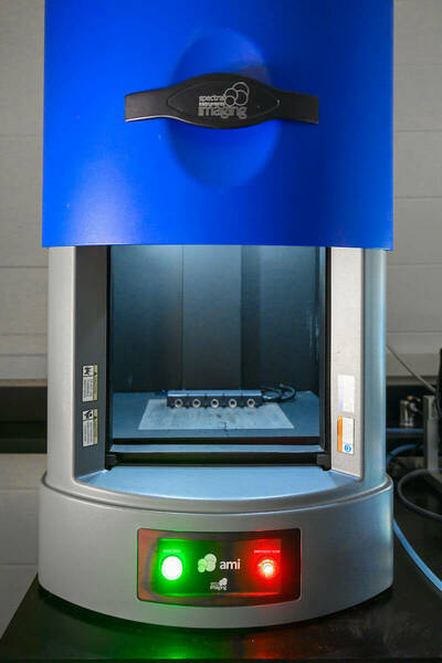
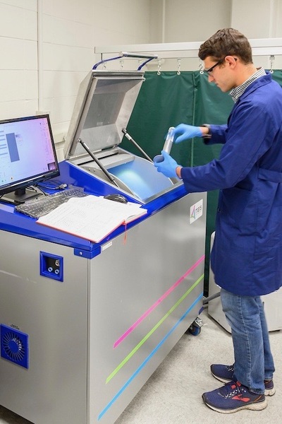
Advanced Molecular Imaging HT Instrument
Small animal imaging platform capable of capturing luminescent and fluorescent images via a large aperture lens with automation for filter and field of view selection
-
Body Composition Analysis device for live animals, measuring fat, lean, water and bone mineral content
-
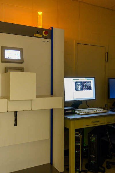
Scanco VivaCT 80 Micro X-ray CT
In-vivo microCT scanner offering the largest field of view (80mm) and the best resolution (5µm)
-
The multiphoton microscope STELLARIS 8 DIVE provides you with flexible multicolor imaging beyond 1 mm in depth for intravital live animal imaging.
Histology Core
The Histology Core provides a means to examine biological processes in mice, rats, frogs, zebra fish, fruit flies, sheep bone, and even human tissue by use of immunohistochemical techniques and pathology.
-
Leica TP 1020 Tissue Processor
Tissue Processor with pre-heat and pre-cool capabilities to maintain constant temperature
-
Embedding station which enables the user to produce paraffin embedded tissue blocks
-
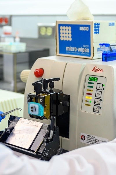
Leica AUTOCUT Microtome and Waterbath
The Leica HistoCore AUTOCUT is developed for fully motorized sectioning of paraffin and plastic-embedded sections
-
The Leica CM1850 Cryostat has proven to regarded as one of the most reliable cryostats in the industry
-
Sample Preparation and Developed Staining Protocols
The staff of the NDIIF have a library of existing protocols and will also work alongside the user to develop and optimize new protocols for sample preparation and staining
-
The fully-automated Leica VT1200 S is recommended for multi user laboratories where users of both semi-automated vibrating blade microtomes and fully automated instruments work together.
-
The CryoJane Tape-Transfer System for Cryosectioning makes it easy to produce high-quality frozen sections of difficult tissues. The CryoJane uses adhesive coated slides and adhesive tapes to capture sections instead of using an anti-roll plate or brush.