Guest Speaker at MWIM Workshop at Notre Dame on May 8th, 2018 at McKenna Hall.
To register and to see an entire list of speakers and the agenda visit imaging.nd.edu
Dr. MacLaren's research concentrates on using electron microscopy to study the structure and chemistry of materials and devices at the nanoscale and below. His group is interested in the link between a material's structure and its function- for example, how the precise arrangement of atoms in a complex oxide can imbue desirable magnetic properties, or how the chemistry of nanometer-sized precipitates enhances the strength of steels.
Current research includes projects on:
- The atomic structure of nanoscale features such as boundaries and defects in functional oxide materials
- The atomic structure of dielectric optical coatings for high precision interferometry (in collaboration with the Institute for Gravitational Research
- The structure and chemistry of nanoprecipitates in steels
- The nanoscale mechanisms of corrosion of Zircaloy in pressurized water
- Absolute quantification of the nanoscale chemistry of materials
- Novel developments in the use of fast pixel detectors for scanning transmission electron microscopy
- Quantitative studies of the structure and chemistry of semiconductor heterostructures, especially for optoelectronic devices
Some of Dr. MacLaren’s publications include:
Craven, A. J., Sala, B., Bobynko, J. and MacLaren, I. (2018) Spectrum imaging of complex nanostructures using DualEELS: II. Absolute quantification using standards. Ultramicroscopy, 186, pp. 66-81.(doi:10.1016/j.ultramic.2017.12.011)
Bashir, A. et al. (2018) Interfacial sharpness and intermixing in a Ge-SiGe multiple quantum well structure. Journal of Applied Physics, 123(3), 035703. (doi:10.1063/1.5001158)
Peng, H., Wang, G., Wang, S., Chen, J., MacLaren, I. and Wen, Y. (2018)Key criterion for achieving giant recovery strains in polycrystalline Fe-Mn-Si based shape memory alloys. Materials Science and Engineering A: Structural Materials Properties Microstructure and Processing, 712, pp. 37-49. (doi:10.1016/j.msea.2017.11.071)
Annand, K., Nord, M., MacLaren, I. and Gass, M. (2017) The corrosion of Zr(Fe, Cr)₂ and Zr₂Fe secondary phase particles in Zircaloy-4 under 350ºC pressurised water conditions. Corrosion Science, 128, pp. 213-223. (doi:10.1016/j.corsci.2017.09.014)
Mir, J.A. et al. (2017) Characterisation of the Medipix3 detector for 60 and 80 keV electrons. Ultramicroscopy, 182, pp. 44-53.(doi:10.1016/j.ultramic.2017.06.010)
He plays a leading role in the Materials and Condensed Matter Physics Group at the University alongside the group leader, Dr Stephen McVitie. He is heavily involved in the running of the Kelvin Nanocharacterisation Centre- a Glasgow facility combining TEM/STEM facilities with Focused Ion Beam, Scanning Probe Microscopy, Conventional Sample Preparation, and detector development for Electron Microscopy, and providing nano- and micro-scale characterization services to an interdisciplinary range of users spanning many fields of Engineering, Chemistry, Earth Sciences and Life Sciences.

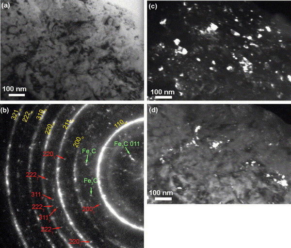
Fig. 1. TEM images of the microstructure of the steel after HPT deformation: (a) bright-field image; (b) zero-loss filtered SAD pattern, ferrite rings are labelled and indexed, some cementite reflections are indicated, and austenite reflections are indexed; (c) dark-field image using the 1 1 0 reflection of ferrite; (d) dark-field image created using some of the cementite reflections. (Fig. (b,c) are reprinted from Ref. [20] with permission of WILEY-VCH.)