
Hafnia (HfO2) nanoparticles as an Xray contrast agent and midinfrared biosensor
T.L. McGinnity, O. Dominguez, T.E. Curtis, P.D. Nallathamby, A.J. Hoffman, R.K. Roeder, Nanoscale. 2016, 8, 13627–13637. DOI:10.1039/C6NR03217F.
Hafnium oxide (HfO2) nanoparticles (NPs) possess unique functional properties for use as an X-ray contrast agent and mid-infrared biosensor. Imaging phantoms comprising HfO2 NPs, Au NPs (used in preclinical research), and iodine (used in clinical medicine), were imaged using a novel photon counting, preclinical spectral CT to enable multi-energy imaging and exploit energy-dependent differences in the X-ray attenuation of the three contrast agents. Spectral CT enabled simultaneous color discrimination and quantitative molecular imaging of the HfO2 NPs, Au NPs, and iodine contrast agents, which is not possible using conventional CT due to relatively similar overall attenuation. Contrast agents were also able to be clearly distinguished from bone and soft tissue (water).
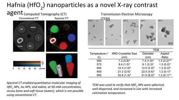
Transmission electron microscopy was used to verify the preparation of spherical HfO2 NPs of controlled size by a solgel process. Control of the calcination temperature allowed the NP size to be tailored over a fourfold range of ~731 nm at 500-950°C. Importantly, NPs in this size range are suitable for in vitro labeling, in vivo delivery, and cellular internalization. Thus, the work in this paper utilized both the in vivo imaging and electron microscopy facilities in the NDIIF to characterize the functional properties and nanostructure, respectively, of novel nanoparticles.