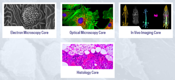
The Notre Dame Integrated Imaging Facility has been embedded within the University of Notre Dame campus for over ten years working closely with students and faculty alike at the forefront of research to develop and explore critical questions that create and explore opportunity for health, innovation, and advancement.
2019 was a year of re-focusing, re-defining, and re-building.
The arms of the NDIIF touch many areas of research on our campus and beyond. Our researchers focus their efforts on nano-materials, conductivity energy, biological development and functionality, chemical analysis and reactivity, and disease and distribution among many others. Over the past decade hundreds of individuals have passed through our facilities collecting images and data that is dispersed and recognized world-wide. As expected, our instruments have started to display their wear and tear. In early January, the NDIIF worked to replace our Helios FIB/SEM instrument with a 4th generation extreme high resolution field emission scanning electron microscope with focused ion beam technology. This instrument offers simultaneous imaging and patterning with end-point detection through real-time monitoring. This instrument provides the most advanced focused ion- and electron beam performance with resolution of 5nm and 0.6nm respectively. The user is able to achieve greater milling speed without sacrificing quality for large areas and repetitive milling projects. It allows the industry's best image resolution for quick identification and metrology of thin layers and substructures as well as other overall productivity improvements.
We have re-defined our services by introducing a new team that provides expert instruction to train and develop users of all levels. In June of 2019, Maksym Zhukovskyi transitioned into the role of TEM program director. Maksym’s level of knowledge and experience throughout his time here on our campus as a PhD candidate and post-doctoral fellow, allowed him the confidence and skills necessary to easily transition into a leadership role. His footprint in the field of electron microscopy is driven by energy and passion to explore new capabilities within the field and the ease of meeting researchers along their career path to offer insight and direction in territories previously uncovered and those that remain still hidden.
Our capabilities within the NDIIF were re-defined by adding new tools and techniques for sample preparation and analysis. The histology core has successfully developed SEM sample preparation protocols and worked with nearly a dozen researchers to prepare cells, bacteria and nanomaterials for imaging using SEM instruments. Additionally, the histology core has worked hand in hand with the electron microscopy core to implement TEM sample preparation using a new ultramicrotome. A series of instruments were acquired by Jennifer Schafer and a team of faculty through the Universities equipment renewal and replacement fund. This ERRP funding allowed the NDIIF the ability to acquire a new ultramicrotome that is capable of sectioning both room temperature samples embedded in resin or plastic as well as cryo-perserved samples that must be sectioned at sub-zero temperatures. In addition, the NDIIF worked closely with researchers to acquire a cryo-plunger that is able to quickly and efficiently flash freeze a sample of soft material or biological material in less than a second that can then be directly transferred to the TEM equipment for imaging. Maksym has been instrumental in working with researchers to develop and define imaging protocols to satisfy the goals of the experiment and produce numerous high quality images.
In January of 2019, the optical microscopy core through the hiring of Dr. Katharine White was able to acquire a Nikon spinning disk microscope. This microscope is housed on the first floor of Harper Hall and is available to all researchers and students throughout the campus of Notre Dame and our CTSI partners. The Nikon CSU X1 confocal with solid-state high-power laser launch, motorized filter wheel, and powerful sCMOS camera is a precision optical instrument. This system is faster, brighter and more efficient allowing for multimode imaging of both live and fixed samples using lower power lasers and shorter exposure times. The spinning disk system is coupled with an inverted Ti2 base allowing users to perform confocal, photoactivation and widefield imaging in the same experiment.
New analysis tools were added to the NDIIF to improve and enhance the amount of data that one could obtain from a single image or a series of images. IMARIS is the world-leading interactive microscopy analysis software used by microscopists around the world. This data analysis software package provides machine-learning tools for clarification and identification of spots and surfaces within the image to improve statistics and enhance data interpretation within an image. This powerful software is a full service option for analyzing images of bioimaging and life science, electron microscopy, fluorescence microscopy, x-ray fluoroscopy, radiography, and CT images! Users have the ability to use the software by visiting the offline computer station or logging in remotely through a computer platform elsewhere.
The opportunity to re-focus and re-define the NDIIF was quickly brought to a halt in March of 2020 when the world was faced with a pandemic that we still continue to address. Although this halt in research efforts may have seemed a bit debilitating at times, the NDIIF saw this as an opportunity to rebuild and expand our efforts to continue to support researchers and our community.
During our time away from campus the NDIIF team met with researchers to discuss the purchase of a new light sheet microscope. Funding for this instrument was earned in partnership with Dr. Jeremiah Zartman, Brad Smith, Cody Smith, Pinar Zorlutuna, and Holly Goodson through a NSF written and awarded grant. A new Bruker Luxendo Multiple-View Selective-plane is currently being installed on the campus of Notre Dame. This system provides four orthogonal views of the sample with no need for rotation, maximized acquisition speed, long-term stability of the sample and high precision of data fusion. This system will be set up in room 010b of Galvin Hall and will be available to researchers throughout the campus and to our extended CTSI partners.
In April of 2020 when we all were settling into our new work environments, Sara Cole was settling into a new home within Indiana. Sara comes to us from the Ohio State University where she was their previous Associate Director of Microscopy. She joined the NDIIF team on April 1 and is serving as our Optical Microscopy Program Director. Her expertise and publication record are outstanding and will offer the University a great deal of opportunity and innovation. As the program director Sara will coordinate training and educational outreach programs, develop and implement protocol improvements and maintain as well as work to expand the NDIIF optical microscopy program here on our campus. Her expertise and publication record is outstanding and will offer the University opportunity and innovation. Immediately after Sara’s arrival, she jumped on board and worked with a large team of faculty to write a NIH shared instrumentation grant. This grant seeks to earn funding to purchase two new instruments that will fill in gaps between the optical microscopy core and the InVivo imaging core. In addition, Sara was chosen to submit a Chan Zuckerberg Initiative Imaging Scientist application. If funded, this will significantly improve the core’s ability to enhance our remote training and education programs. The future is bright with Sara on our team!
Finally our efforts to rebuild have allowed us to truly evaluate the lifetime of our instruments and make decisions regarding whether we should invest in replacing or renewing our current instruments. This will allow us to remove unused equipment to make space and provide resources for purchasing newer equipment that will continue to serve our existing users as well as increase capabilities to improve our research offerings. In partnership with centers and institutes across the campus such as the Harper Cancer Research Institute and Friemann Life Science Center, the University is able to offer facilities such as the Pre-Clinical Therapeutics Facility. In addition, the NDIIF is partnering with other cores to develop new shared research spaces such as the facility now being constructed in the basement of McCourtney Hall. In this space will be housed an environmental SEM and a Near IR Raman Spectroscopy Instrument. But the greatest partnership that is near and dear to the hearts of the NDIIF team is the partnership with our students. Each year we recognize the efforts of our students by presenting an annual imaging award for use of our Biological Imaging Equipment and Electron Beam Equipment as well as what we call “beautiful art” created in science.
This year the winner of our Best Biological Imaging Publication went to Cynthia Spires for her work in Paired Agent Fluorescence Imaging of Cancer in a Living Mouse Using Pre-assembled Squaraine Molecular Probes with Emission Wavelengths of 2690 and 830nm featured in Bioconjugate Chemistry.
The winner of our Best Electron Beam Imaging Publication went to Spencer Golze for his work in Plasmon Mediated Synthesis of Period Arrays of Gold Nanoplates Using Substrate-Immobilized Seeds Lined with Planar Defects featured in NanoLetters.
And to recognize the beauty that is often seen but not always present in publications, we awarded the Best Artistic Image to Brooke Chambers. Her work featured a reflected 10X image of a beautiful model organism, the Danio rerio embryo, commonly known as the zebrafish. You can read more about each one of these awards on our website and our LinkedIN channel. Congratulations to all of the nominations for excellence in imaging!
For the past 10 years, through the world pandemic and for the next decades to come… The Notre Dame Integrated Imaging Facility is HERE for you.