Post-Translational Modification of the Membrane Type 1 Matrix Metalloproteinase (MT1-MMP) Cytoplasmic Tail Impacts Ovarian Cancer Multicellular Aggregate Dynamics
Yang, J., Kasberg, W. C., Celo, A., Liang, Z., Quispe, K., & Stack, M. S. (2017). Journal of Biological Chemistry, 292(32), 13111-13121.

The combination of several techniques including the use of optical light microscopy and scanning electron microscopy proved to reveal hidden information that is deemed remarkable in the field of cancer research. Research done by Sharon Stack and her colleagues divulged significant information regarding membrane type 1 matrix metalloproteinase (MT1-MMP, MMP-14), a transmembrane collagenase highly expressed in metastatic ovarian cancer. Accumulating evidence shows that the cytoplasmic tail of MT1-MMP is subjected to phosphorylation, and this post-translational modification regulates enzymatic activity at the cell surface. To investigate the potential role of MT1-MMP cytoplasmic residue Thr567 phosphorylation in regulation of metastasis-associated behaviors, ovarian cancer cells that express low endogenous levels of MT1-MMP were engineered to express wild-type MT1-MMP, a phosphomimetic mutant (T567E), or a phosphodeficient mutant (T567A). To examine the potential impact of MT1-MMP on MCA dynamics, EOC MCAs were produced using the hanging drop method from cells expressing wild-type MT1-MMP or Thr567 mutants and were evaluated by light and scanning electron microscopy (SEM) to assess aggregate area and overall morphology.
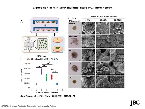
Similar to light microscopic observations, examination by high-resolution SEM shows loosely clustered aggregates of cells in OVCA433 MCAs. Additionally, the electron microscope offers atomic resolution capabilities and is able reveal characteristics that otherwise might have gone unnoticed. The data acquired from these and other corresponding studies is remarkable and will catapult our research into a new era of discovery. The ability to combine the expertise of each of these imaging fields allows us to advance our goal of increasing the survival rate of women with ovarian cancer. This is the third time that Sharon Stack’s work has been featured as a cover story.