Rajesh Sahadevan
2014 Best Electron Microscopy Publication Imaging Award
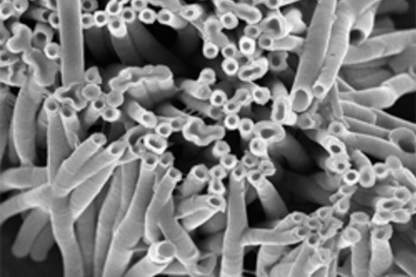
The 2014 Best Electron Microscopy Imaging Publication was awarded to Rajesh Sahadevan, a post-doctoral research associate with Professor W. A. Phillip in in the Department of Chemical and Biomolecular Engineering. Sahadevan and coworkers published a paper titled “Mixed Mosaic Membranes Prepared by LayerbyLayer Assembly for Ionic Separations”. The project addressed the fabrication of mixed mosaic membranes (MMMs) with discrete positively charged and negatively charged domains. Due to this unique nanostructure, MMMs are capable of selectively separating dissolved ions from similarly-sized neutral solutes.
Using the Magellan 400 scanning electron microscope (SEM) located in Stinson Remick Hall, micrographs were acquired that illustrated the feasibility of a layer-by-layer (LbL) technique to fabricate these unique membranes. Furthermore, SEM micrographs of the polymeric nanotubes helped control the dimensions (i.e., length, inner diameter and outer diameter) of these building blocks. The study was published in ACS Nano, 2014, 8, 12338–12345, DOI: 10.1021/nn504736w.