Helios G4 Ux Dual Beam
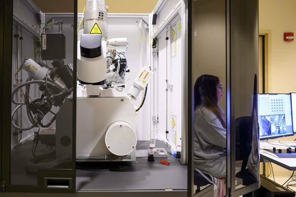
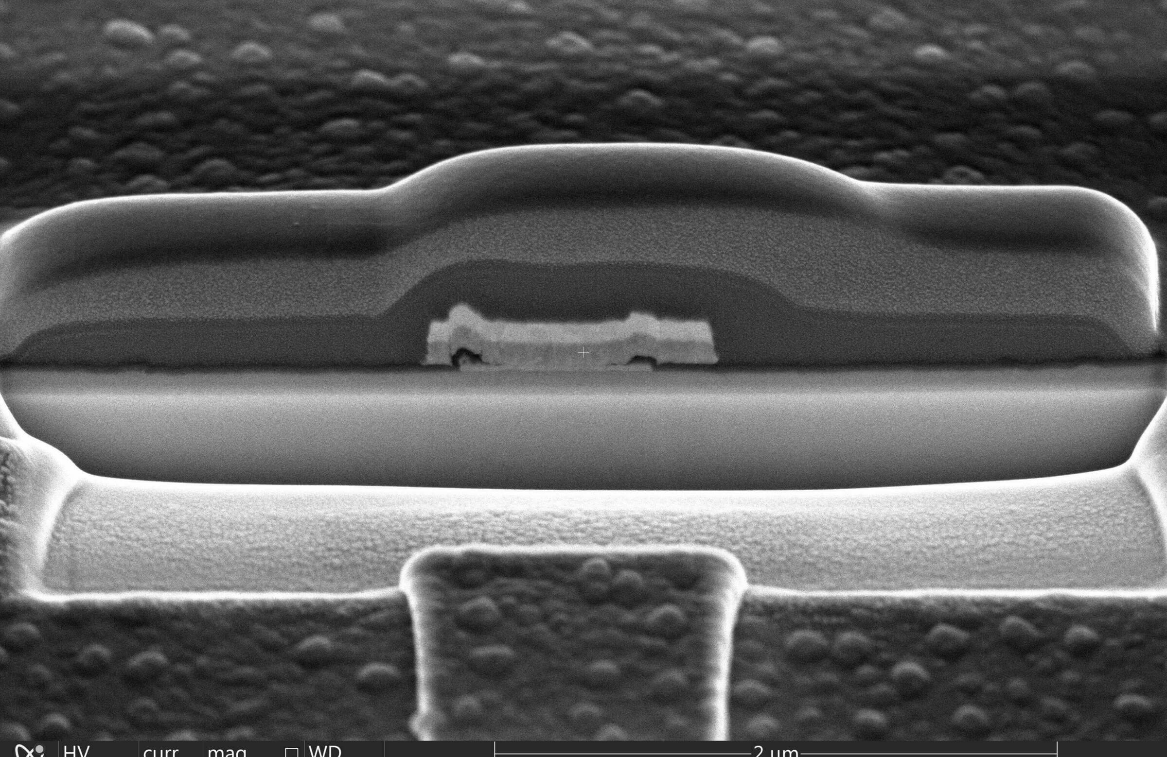
Helios G4 UX Dual Beam (electron & ion beams) microscope is designed for the highest-quality imaging, ultra-thin TEM and APT sample preparation, milling and deposition of complex structures with critical dimensions of less than 10 nm, multi-modal subsurface and 3D data collection.
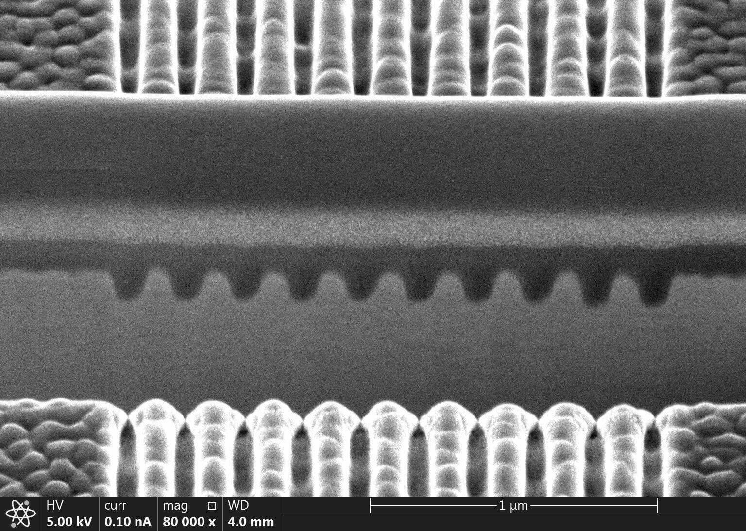
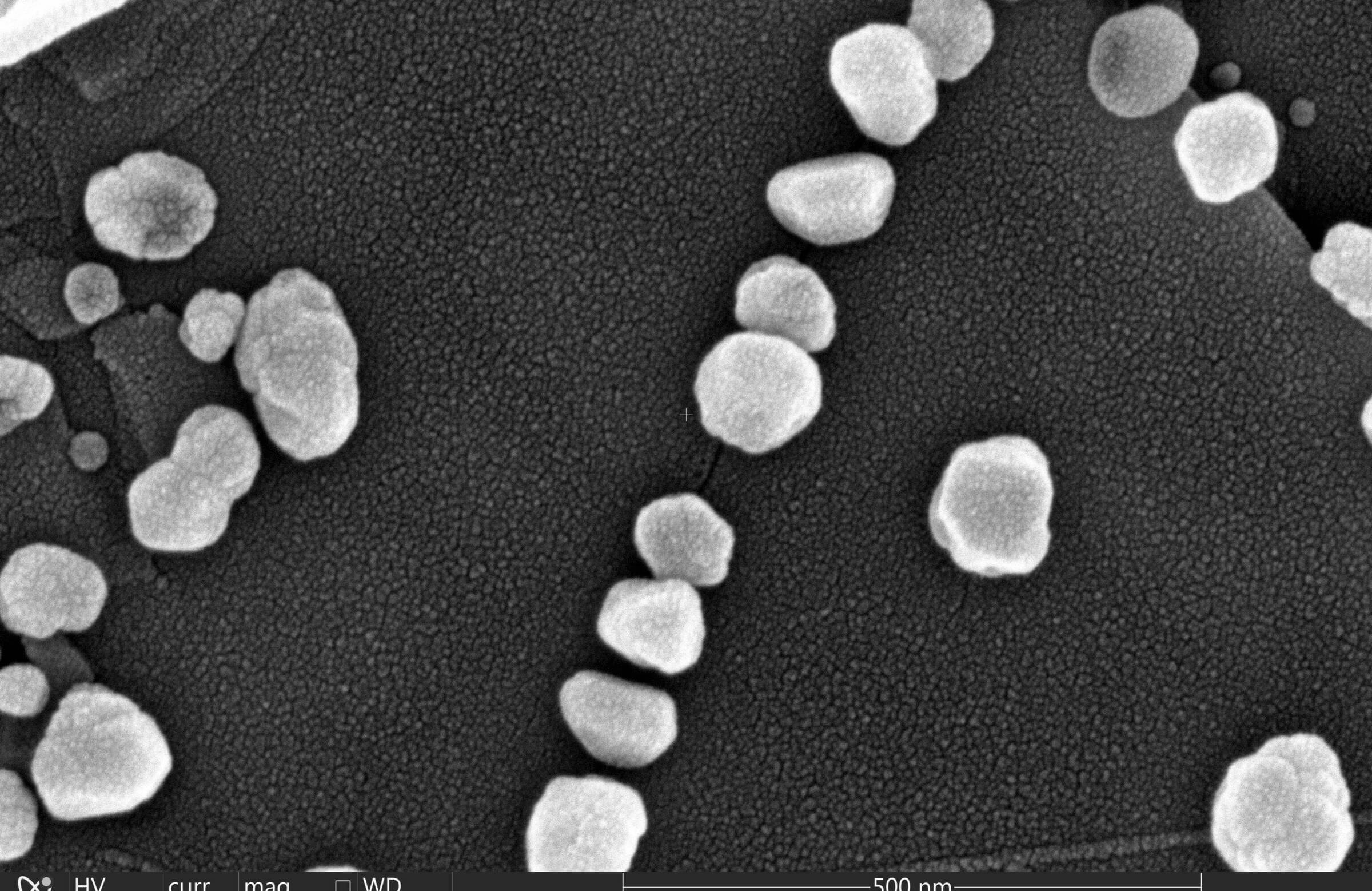
- Electron beam imaging provides the most complete sample information with sharp, refined, and charge-free contrast obtained from 5 integrated in-column and below-the-lens detectors.
- Reveal the finest details with the next-generation UC+ monochromator technology with higher current, enabling sub-nanometer performance at low energies
- The highest quality, multi-modal subsurface and 3D information with the most precise targeting of the region of interest using optional Auto Slice & View™ 4 (AS&V4) software.
- Precise sample navigation tailored to individual application needs thanks to the high stability and accuracy of the 150 mm Piezo stage and in-chamber Nav-Cam.
- Artifact-free imaging based on integrated sample cleanliness management and dedicated imaging modes such as SmartScan™ and DCFI (drift correction frame integration).
Features & Specifications
Electron optics
- Source: Schottky field emitter
- Resolution: 0.6 nm at 15 – 2 kV 0.7 nm at 1 kV (with Beam Deceleration) 1.0 nm at 500 V (with Beam Deceleration and ICD)
- Acceleration Voltage: 350 V to 30 kV
- Landing energies: Adjustable from 20 eV to 30 keV
- Beam current: 0.8 pA – 100 nA
- Dual-mode magnetic immersion / field free lens electron optics
- Everhart-Thornley SE detector for SE detection
- In-lens SE and BSE detection specially designed for high-resolution imaging at both high and low kV’s;
- Mid-column (MD) and In-column (ICD) backscattered electron detectors
- Retractable BS detector, designed for Z- contrast imaging
- UC+ monochromator technology with higher current, enabling sub-nanometer performance at low energies
- Beam deceleration technology for low energy imaging of non-conductive specimens;
Ion optics
- Phoenix field emission focused ion beam optics with liquid Gallium ion emitter.
- Source lifetime: 1000 hours
- Voltage: 500 V to 30 kV
- Beam current: 0.1 pA - 65 nA (15-position aperture strip)
- Resolution: 4.0 nm at 30 kV
- Detection: Everhart-Thornley and ICE detectors ( secondary electron and ion detectors)
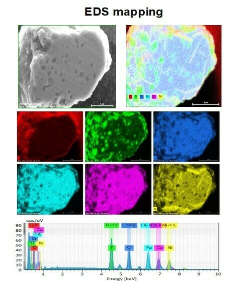
Energy Dispersive X- Ray Spectrometer (EDS )Bruker
- SLEW Window, 60mm2, detection of Boron and up;
- Energy resolution 129 eV (MnKα);
- Elemental mapping, line scan, point analysis.
Location
Stinson Remick B28