Optical Microscopy Core
The Optical Microscopy Core provides researchers with the opportunity to acquire informative single images and three-dimensional reconstructions of fluorescently labeled cells and tissues, either fixed or live.
-
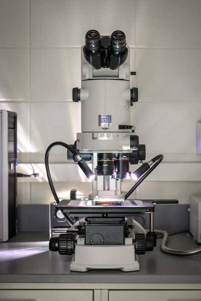
The Nikon AZ100 is a multi-zoom macroscope with a wide range of magnification settings (5x-400x) used for either brightfield (top lit or backlit) or contrast imaging
-
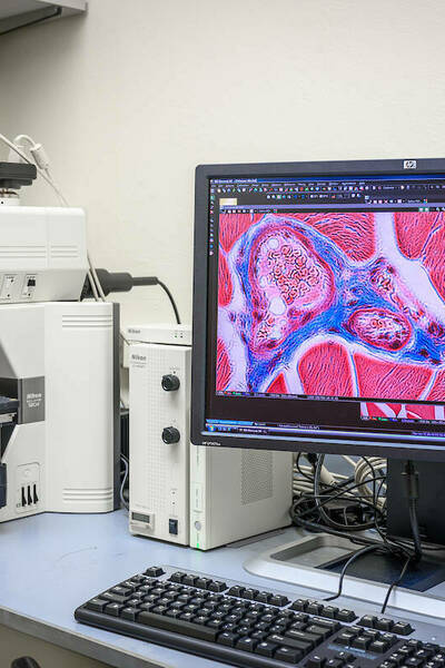
Nikon Eclipse 90i Widefield Fluorescent Microscope
The 90i is useful for imaging fluorescent or chromogenic (chemically stained) samples such as H&E, immunohistochemistry (i.e. DAB, HRP), Gram, trichrome, and more
-
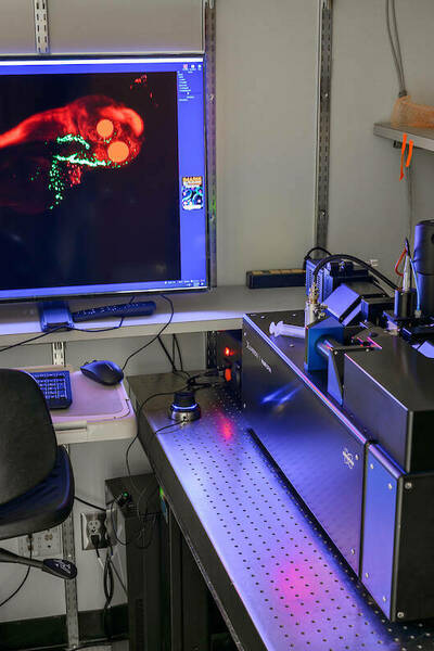
Bruker Luxendo MuVi SPIM Light Sheet
The lightsheet system is ideal for long-term, three-dimensional fluorescence imaging of organisms, tissue explants, and 3D cell culture systems. This microscope can be used for live, fixed, and optically cleared samples
-
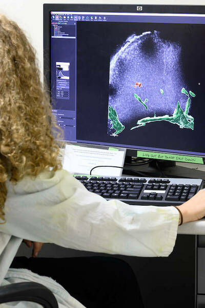
IMARIS software can be used for image presentation and/or data analysis from a variety of commercial fluorescence microscopy image file formats
-
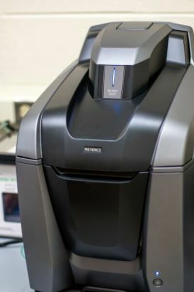
The Keyence BZ-X810 is a fully automated, brightfield and fluorescence microscope adaptable to many different research needs.
-
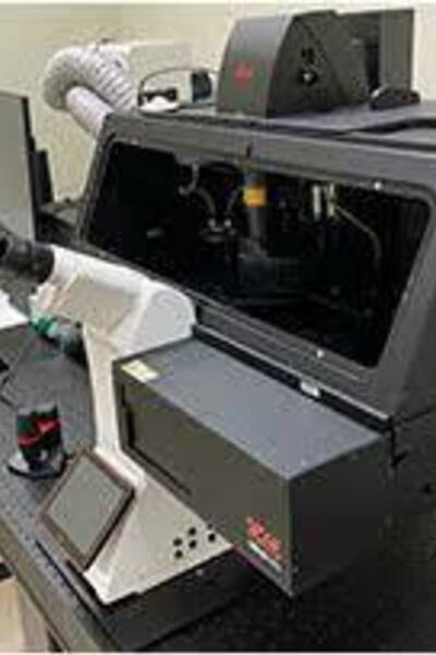
The multiphoton microscope STELLARIS 8 DIVE provides you with flexible multicolor imaging beyond 1 mm in depth.
-

The Zeiss LSM 980 is designed to meet the evolving demands of cutting-edge research, offering advanced imaging capabilities for a broad range of sample types, including organoids, spheroids, single-cell resolution, and real-time dynamic processes.
-
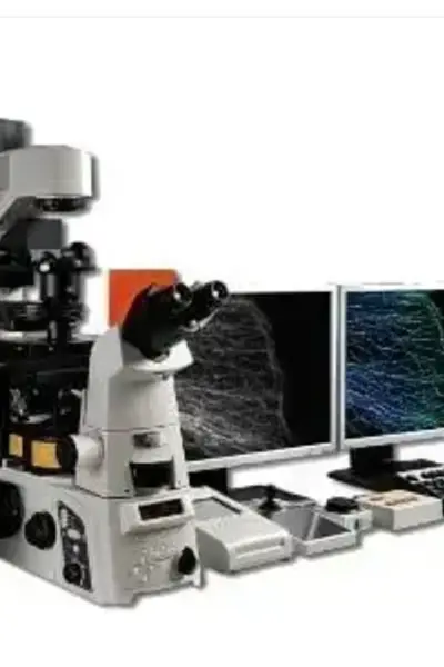
The Nikon N-STORM (STochastic Optical Reconstruction Microscopy) is a super-resolution optical microscope system based on an inverted research stand (Eclipse Ti2-E / Ti-E). It uses single-molecule localization microscopy (SMLM) to bypass the diffraction limit, delivering a 10-fold improvement in resolution over standard optical systems.