- Home ›
- Services ›
- Histology Core ›
- Sample Preparation and Developed Staining Protocols
Sample Preparation and Developed Staining Protocols

Features & Specifications
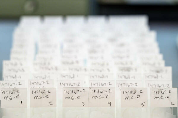
- Staining of tissue sections ranges from routine Hematoxylin-Eosin (H&E's) to special stains demonstrating specific tissue structures.
- Masson’s Trichrome, Periodic Schiff, and Verhoff Van Geison among other special stains are developed and provided as needed.
- The Histology Core is also able to offer a variety of specific immunohistochemical stains.
- Histopathologic interpretation of tissues is available.
- Electron microscopy samples can be prepared either by critical point druing or resin-embedding wthin the histology core as well.
